


Figure 2. ACE2 protein is detected in human dorsal root ganglia using the abcam ab15348 antibody.
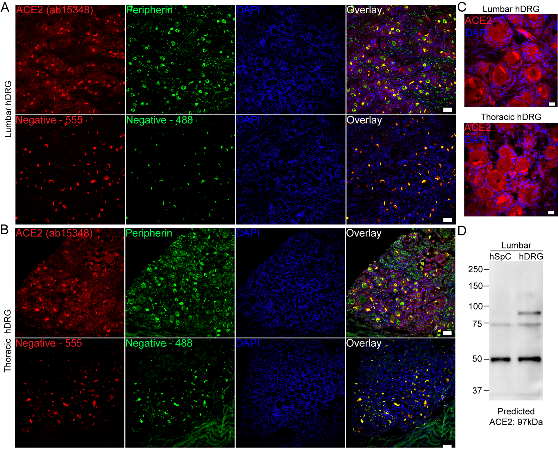
A) Representative 10x images of protein staining for ACE2 (red; Abcam ab15348), peripherin (green; a sensory neuron marker) and DAPI (blue) in lumbar and B) thoracic human dorsal root ganglia. C) 40x images showing ACE2 signal in both neurons and non-neuronal cells including satellite glial cells. D) Western blot of ACE2 using ab15348 on lumbar spinal cord and DRG revealed a strong band at the predicted molecular weight of ACE2 protein in human DRG but not spinal cord, and other non-specific bands. Scale bars: 10x = 100μm, 40x = 20μm.