
Figure 17.
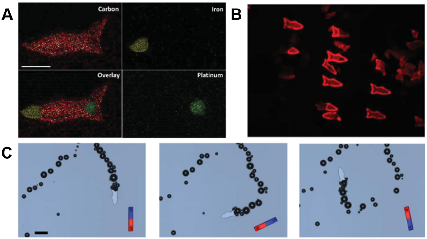
3D printed microfish. (A) Energy-dispersive X-ray spectroscopy showing 3D microfish with different nanoparticles localized at the head, tail, and body. (B) Fluorescent image of the microfish after detoxification of a melittin solution. (C) Time-lapse images of the microfish performing sharp turns with magnetic guidance. (A–C) Reproduced with permission from ref 280. Copyright 2015 Wiley-VCH.