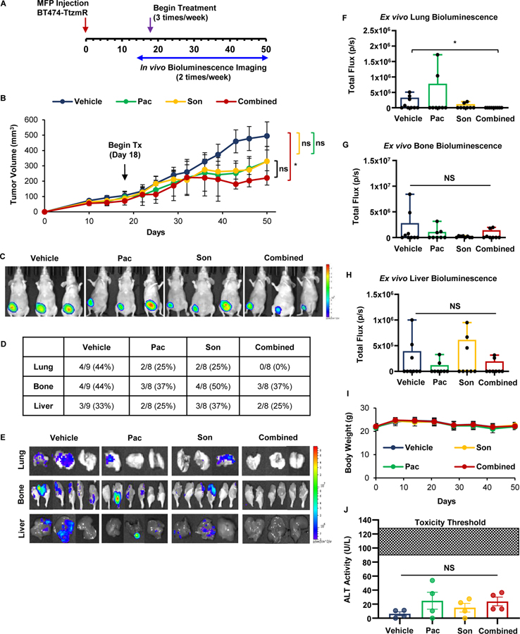
Figure 5: Co-treatment with JAK2 and SMO inhibitors suppresses the orthotopic growth and lung metastasis of HER2-positive/trastuzumab-resistant breast cancer in vivo.
(A) Schema for MFP mouse model. Luciferase-expressing BT474-TtzmR breast cancer cells were implanted into the right inguinal MFP. Tumor growth was assessed via caliper measurements and bioluminescent imaging. Once tumors reached an average of 90 mm3, mice were randomized into vehicle, 7.5 mg/kg pacritinib, 20 mg/kg sonidegib, or combination treatment groups (N=8–9) and received three intraperitoneal treatments per week. (B) Growth curve for MFP tumors. (C) Representative bioluminescent images of xenograft-bearing female nude mice 50 days post-inoculation. (D) Quantification of organ-specific metastasis incidence. (E) Representative bioluminescent ex vivo organ images are shown. (F–H) Ex vivo lung (F), bone (G), and liver (H) bioluminescence was quantified. (I) Average mouse weights over the course of the study. (J) Circulating alanine transaminase (ALT) levels were not significantly increased in any treatment group. Data presented as mean ± SEM. One-way ANVOA with Tukey’s multiple comparison post hoc test was used to compute p values. *, p < 0.05

