Fig. 4.
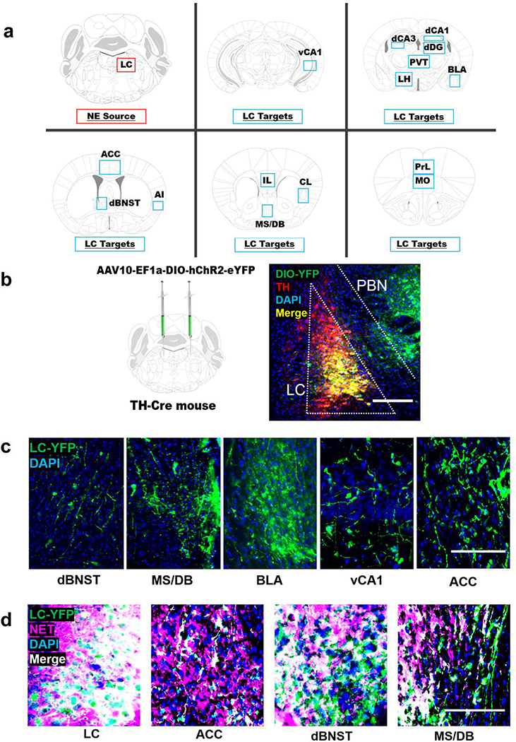
Viral tracing of locus coeruleus (LC) fibers in forebrain regions implicated in novelty. a Anatomical location of the LC and 15 target regions in the forebrain: medial orbitofrontal cortex (MO), prelimbic (PrL) and infralimbic cortices (IL), anterior cingulate cortex (ACC), agranular insula (AI), claustrum (CL), medial septum/diagonal band (MS/DB), dorsal bed nucleus of the stria terminalis (dBNST), basolateral amygdala (BLA), dorsal dentate gyrus (dDG), dorsal hippocampal subfields CA1 (dCA1) and CA3 (dCA3), ventral hippocampus subfield CA1 (vCA1), lateral hypothalamus (LH), and the paraventricular nucleus of the thalamus (PVT). b TH-Cre mice received bilateral intra-LC infusions of a Cre-dependent viral vector encoding ChR2-eYFP, which becomes concentrated in axons and axon terminals and can be used as a tool for projection mapping. ChR2-DIO-YFP expression (green) substantially overlaps with immunostaining for TH (red) in the LC and is largely restricted to TH+ LC neurons, with some expression in TH- cells of the neighboring parabrachial nucleus (PNB). c Representative micrographs of YFP+ terminals from the LC in the dorsal bed nucleus of the stria terminals (dBNST), medial septum/diagonal band complex (MS/DB), basolateral amygdala (BLA), the CA1 subfield of the ventral hippocampus (vCA1), and the anterior cingulate cortex (ACC). d All YFP+ cell bodies in the LC and most YFP+ terminals in the ACC, dBNST, and MS/DB overlap with immunostaining for the norepinephrine transporter (NET), indicating that these axon terminals originate from LC neurons rather than the PBN. The scale bar denotes 100 μM in all micrographs.

