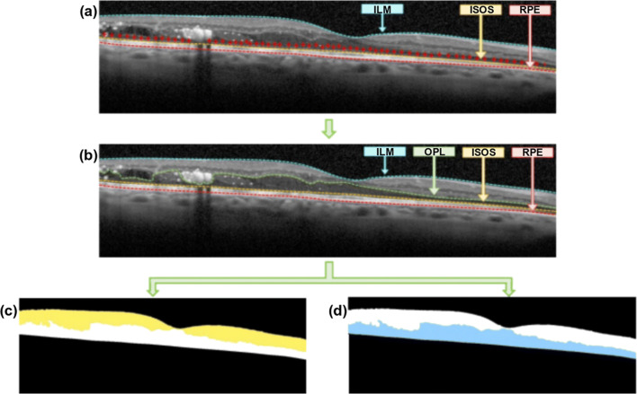
Fig. 4.
Example of OCT image with the identification of the inner and outer retinal regions. a Identification of the ILM, ISOS, and RPE layers. b Additional delimitation of the OPL layer. c Identified inner retina, between the ILM and the OPL layers. d Identified outer retina, between the OPL and the RPE layers

