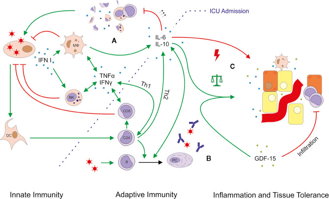
Figure 8.
Simplified outline of immune responses in severe COVID-19 ARDS. Viral infection leads to the production of type I interferon (IFN), which activates macrophages (MФ) and natural killer (NK) cells. Considering a median COVID-19 symptom onset 11 days prior to intensive care unit (ICU) admission, this first-line of defense is likely not depicted by our data. Various mediator cytokines (blue dots) including tumor necrosis factor alpha (TNFα), IFNγ, interleukin (IL)-6 and IL-10 connect innate and adaptive immunity and help to control the pro- and anti-inflammatory response. Severe COVID-19-induced ARDS was accompanied by massively upregulated IL-6, which might explain the observed lymphocytopenia. Excessive levels of IL-6 can block lymphopoiesis (30) and induce lymphocyte death (31). In particular NK cells and CD8+ T cells showed low absolute and relative numbers indicating an impaired or delayed cytotoxic reaction (A). The B cell response and antibody production seemed largely unimpeded (B). Moreover, no obvious imbalance between pro- and anti-inflammatory mechanisms was detected, as IL-10 and growth differentiation factor 15 (GDF-15, green dots) were also upregulated (C). GDF-15 is a member of the anti-inflammatory TGF-β superfamily, which increases the ability of tissues to tolerate inflammatory damage. We thus hypothesize that induction of GDF-15 is an adaptation to restore a disturbed balance between pro- and anti-inflammatory mechanisms. Production, promotion, and positive feedback are illustrated with a green arrow (↑); inhibition and attack is depicted in red (T). The green arrows with the balanced scale represent tissue protection; the red arrow with a lightning symbol depicts inflammation. DC, dendritic cell; PC, plasma cell.

