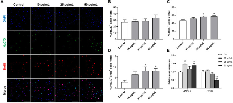
FIGURE 8.
Effects of STEE on proliferation and early differentiation of hNSC. Neurospheres were cultured in proliferation medium with BrdU for 24 h, and BrdU-labeled dissociated cells were visualized by confocal microscopy. (A) Fluorescence images of immunocytochemistry for the neuronal progenitor marker HuC/D and the proliferation marker BrdU in control or STEE-treated cells. Scale bar = 100 μm. (B–D) Quantification of HuC/D, BrdU, or HuC/D/BrdU double-positive cells over total cells visualized with DAPI. n = 8–10 sections per culture. (E) Transcript levels of ASCL1 and HES1 in the cultures at the early differentiation stage. Transcript levels were determined in triplicates from three independent experiments. (B–E) Results are expressed as relative percentages or relative to control values. Values are presented as mean ± SEM. Comparisons were performed using one-way ANOVA followed by Dunnett’s post hoc test: *p < 0.05 and **p < 0.01.

