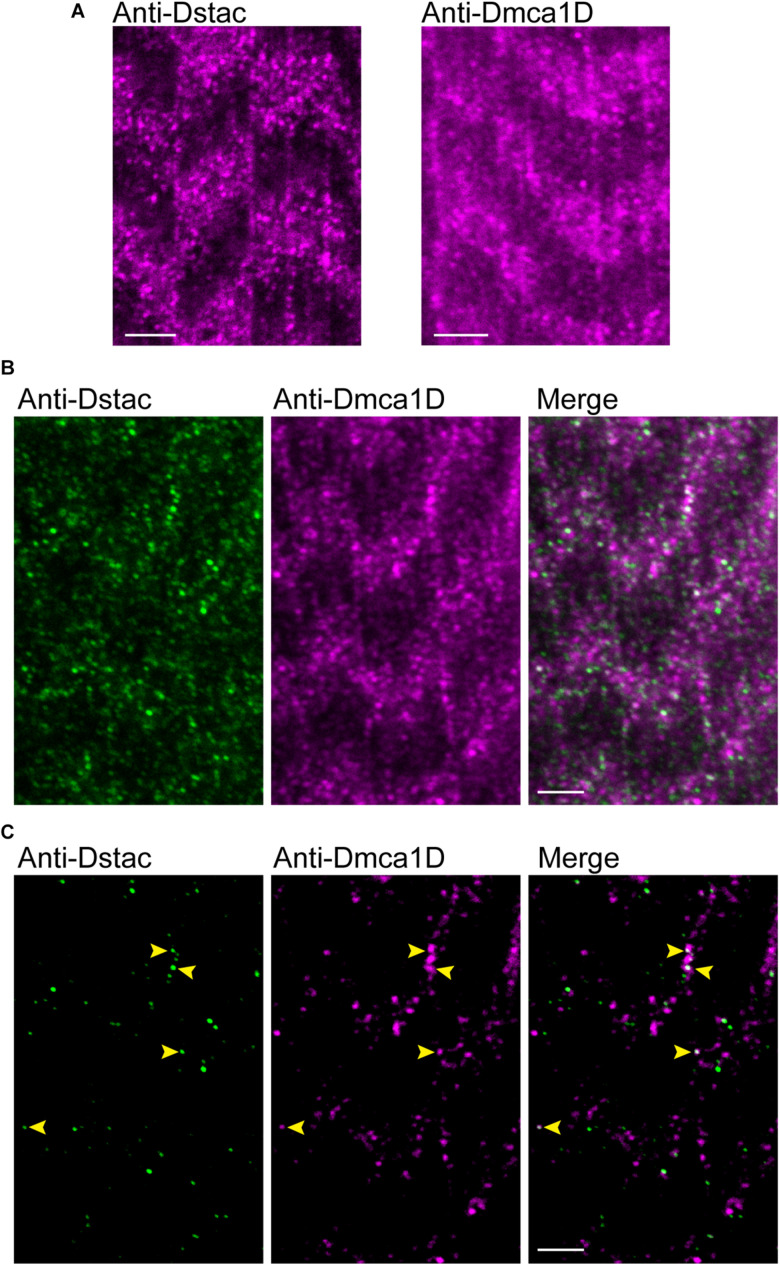
FIGURE 2.
Dstac and Dmca1D are co-expressed in larval muscle stripes. (A) Anti-Dstac (left) and anti-Dmca1D (right) labeling of 3rd instar body-wall muscles showed expression of Dstac and Dmca1D in stripes orthogonal to the longitudinal axis of the muscles. The images are a single focal plane. Scale bar, 3 μm. (B) Co-immunostaining of 3rd instar larval body wall muscles with anti-Dstac and anti-Dmca1D showed co-expression of Dstac and Dmca1D in the same stripes. The images are a single focal plane. Scale bar, 3 μm. (C) Same images as in (B) but showing only the puncta made up of the brightest 50% of pixels for easier comparison of the pattern of anti-Dstac and anti-Dmca1D labeling. Arrowheads indicate some puncta that co-labeled with anti-Dstac and anti-Dmca1D. The images are a single focal plane. Scale bar, 3 μm.

