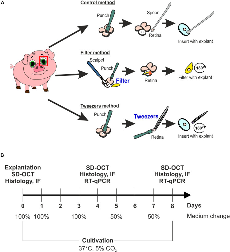
FIGURE 1.
Scheme of the used explant methods and study design. (A) Scheme of the three different explantation techniques, named control, filter, and tweezers method. The fourth group was a native one consisting of samples gained via the three different methods, which were analyzed at day 0. (B) Timeline of the study to investigate which explantation method best preserves the photoreceptor morphology ex vivo. Three techniques were compared during the cultivation periods of 4 and 8 days using spectral domain optical coherence tomography (SD-OCT), immunofluorescence (IF), and quantitative real-time PCR (RT-qPCR). Native samples were also included in the analysis.

