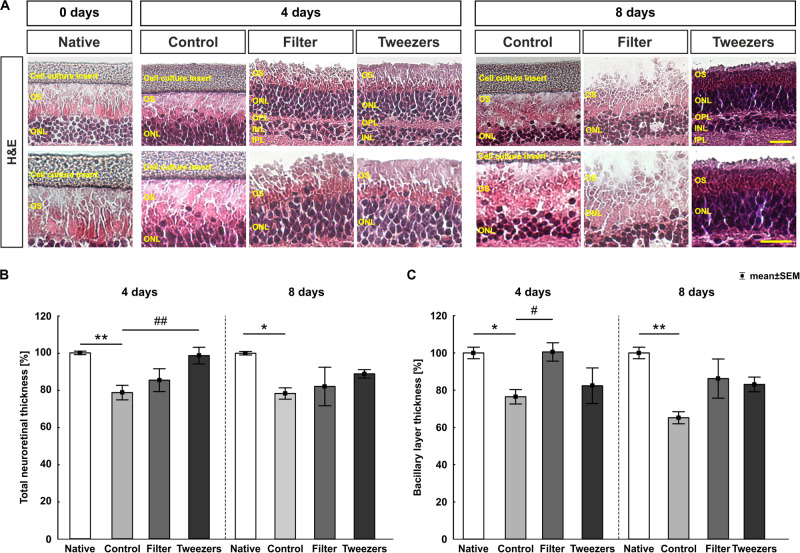
FIGURE 3.
Total and bacillary layer thickness measurement in stained retinas. (A) Representative images of H&E-stained retinas for all three explantation methods and the native group in 400 × (upper panel) and 630 × magnification (lower panel). (B) At 4 days, a significant reduction of the total retinal thickness in control compared to native samples (p = 0.005) was observed, while a preservation was detected in the novel techniques filter and tweezers when compared to native samples. Comparing tweezers to control samples, a significantly thicker retinal thickness was noted (p = 0.009), while a similar thickness was observed between filter and control retinas. Similar results were found at 8 days. A reduction of the total retinal thickness was revealed comparing control to native samples (p = 0.04). A conservation of the total retinal thickness was detected comparing filter and tweezers to native or control retinas. (C) A significant thinning of the bacillary layer was seen in the control group compared to native (p = 0.04) and filter samples (p = 0.046). However, a better-preserved bacillary layer was noted in the tweezers method compared to the native and control samples at 4 days. A reduction in the bacillary layer thickness was measured at day 8 comparing the control and native samples (p = 0.001). The novel methods filter and tweezers maintained the bacillary layer thickness better than did control retinas. OS, photoreceptor outer segments; ONL, outer nuclear layer; OPL, outer plexiform layer; INL, inner nuclear layer; IPL, inner plexiform layer. Scale bars: 20 μm, values are mean ± SEM. n = 9–10/group. *p < 0.05 and **p < 0.01 vs. native group; #p < 0.05 and ##p < 0.01 vs. controls.

