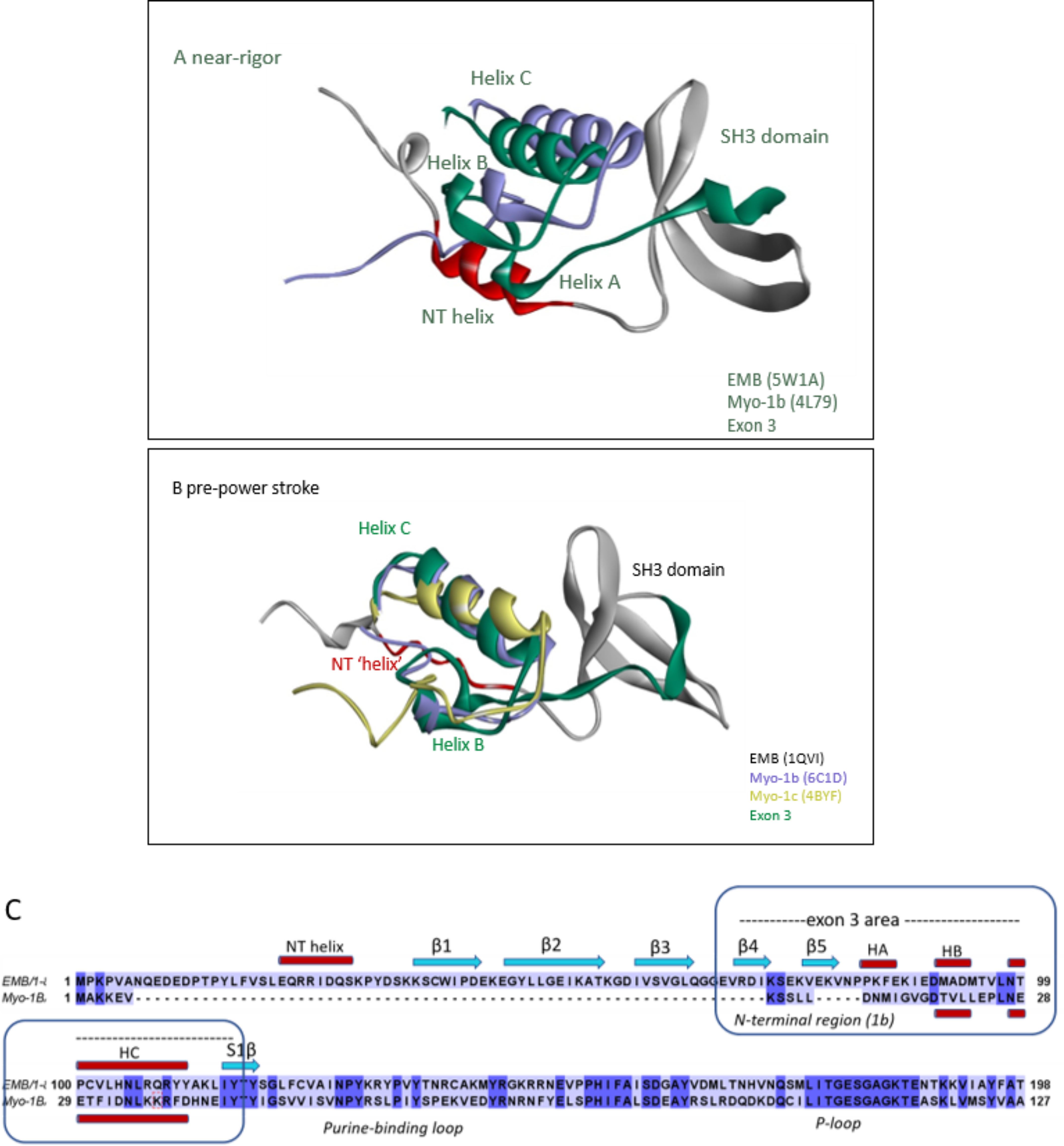
Figure 8.

Overlay of N-terminal regions of EMB and Myo-1b or Myo-1c. A and B, EMB (gray) with the exon 3 area (green) and the N-terminal helix (NT) shown in red, Myo-1b (purple), and Myo-1c (yellow). Note the similar orientation of exon 3 secondary structure elements (helix B and C) of EMB with respect to the NTR of Myo-1b and/or Myo-1c in both near-rigor and pre-power stroke state. A, overlay of crystal structures of Myo-1b (PDB ID: 4L79) with EMB (rigor-like, PDB ID: 5W1A). B, overlay of Myo-1b (PDB ID: 6C1D) and Myo-1c (PDB ID: 4BYF) with EMB homology model (pre-power stroke, PDB ID: 1QVI template). C, sequence alignment of N-terminal regions of EMB and Myo-1b showing conserved secondary structure elements (HB and HC) for the exon-3 encoded region (EMB) and the NTR (Myo-1b).
