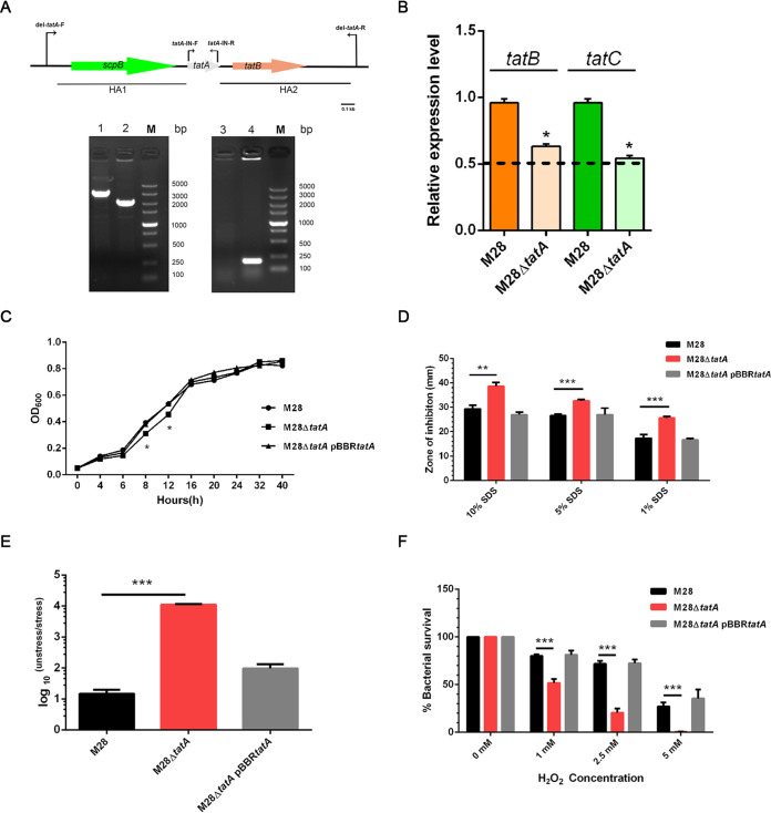
FIG 2.
Mutant construction and growth and sensitivity of various Brucella melitensis M28 strains to stressors. (A) Positions of PCR oligonucleotides used and gel images showing PCR amplicons of the wild type and M28ΔtatA mutant. HA, homologous arm. Lanes: 1 and 2, wild type and M28ΔtatA mutant using del-tatA-F and del-tatA-R primers, respectively; 3 and 4, wild type and M28ΔtatA mutant using tatA-IN-F and tatA-IN-R primers, respectively; M, DNA size marker. (B) Relative transcriptional levels of tatBC in the wild type and M28ΔtatA mutant. Total RNA was extracted from the wild type and the M28ΔtatA mutant, followed by genomic DNA removal, reverse transcription, and qPCR analysis. The expression of target genes in the wild type was normalized to 1, and a 2-ΔΔCt method was employed to indicate expression changes in the mutant. The dashed line denotes a transcriptional reduction of 2-fold. *, P < 0.05. Experiments were repeated 3 times, and values are means ± standard error of the mean (SEM). (C) Bacteria were grown in minimal medium and the optical density was measured at the indicated time points. Data were collected from three replicates, and error bars were too small to be visible on the graph. (D) SDS sensitivity assay. Bacteria (∼1.5 × 10⁷ CFU) were plated onto TSA plates with an SDS-soaked disk placed in the center. The results are plotted as average diameters of the zones of inhibition around the disks. (E) EDTA stress survival assay. Serially diluted cultures of the wild type, M28ΔtatA mutant, and the complemented strain were spotted on plain TSA plates or TSA plates containing EDTA (2 mM). After 72 h of growth at 37°C under 5% CO2, CFU were counted and the ratio of CFU on the TSA plain plates (CFUunstressed) to CFU on the EDTA-supplemented TSA plates (CFUstressed) was calculated for each strain. (F) Survival of bacterial strains at various concentrations of H2O2. Bacterial suspensions containing approximately 5 × 10⁵ CFU were mixed with H2O2 at final concentrations of 5 mM, 2.5 mM, and 1 mM. A control experiment was performed by adding H2O2-free PBS to the bacterial suspensions. After 1 h of treatment at 37°C with shaking, cells were plated onto TSA for counting. The survival of each bacterial strain was determined as the percentage of the control. Experiments were independently performed three times; the data are shown as the mean ± standard error of the mean. Student’s t test was used to evaluate significant differences between the M28ΔtatA mutant and the wild type. *, P < 0.05; **, P < 0.01; ***, P < 0.001.

