FIG 2.
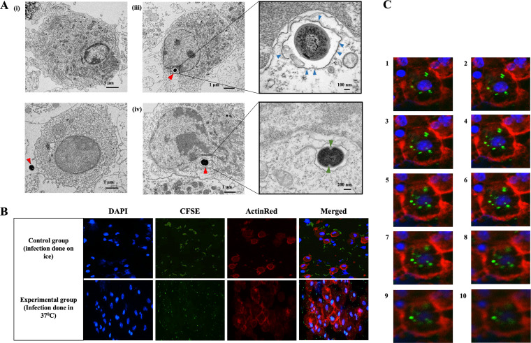
Ultrastructural evidence of bacterial phagocytosis by differentiating and control DCs. (A) TEM images of immature control DC (i) and immature DC with extracellular E. faecalis (red arrow) (iii). (iii, left) E. faecalis engulfed during DC differentiation and found within the cytoplasm. (iii, right) Magnified image of E. faecalis enclosed in a vacuole and surrounded by a single membrane (blue arrows) (iv, left) Intracellular E. faecalis undergoing binary fission. (iv, right) Magnified image showing cell wall invagination (green arrows) that is consistent with cell division. (B) Immunofluorescence staining images showing uptake of CFSE (green)-labeled E. faecalis by control DCs (differentiated in the absence of E. faecalis) (Top) Control DCs incubated on ice, shows E. faecalis outside DCs. (Bottom) DCs at 37°C internalize E. faecalis. (C) Z-stack images, through the DCs, confirming the presence of E. faecalis within the DCs. The experiment was repeated in triplicates three times.

