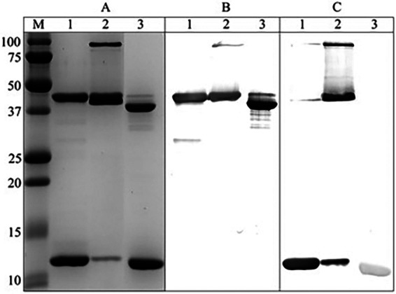
FIG 1.

Characterization of chimera. (A) dsc14CfaE-sCTA2/LTB chimera was separated by 15% SDS-PAGE and stained with Coomassie blue. (B) Immunodetection with CfaE antisera. (C) Immunodetection with LTB antisera. Lanes: M, molecular weight marker (Bio-Rad Precision Plus Protein Standards); 1, heat treated, reduced; 2, unheated, reduced; 3, heat treated, not reduced. Original images of SDS-PAGE gels and blots used to assemble this figure are shown in Fig. S3 in the supplemental material.
