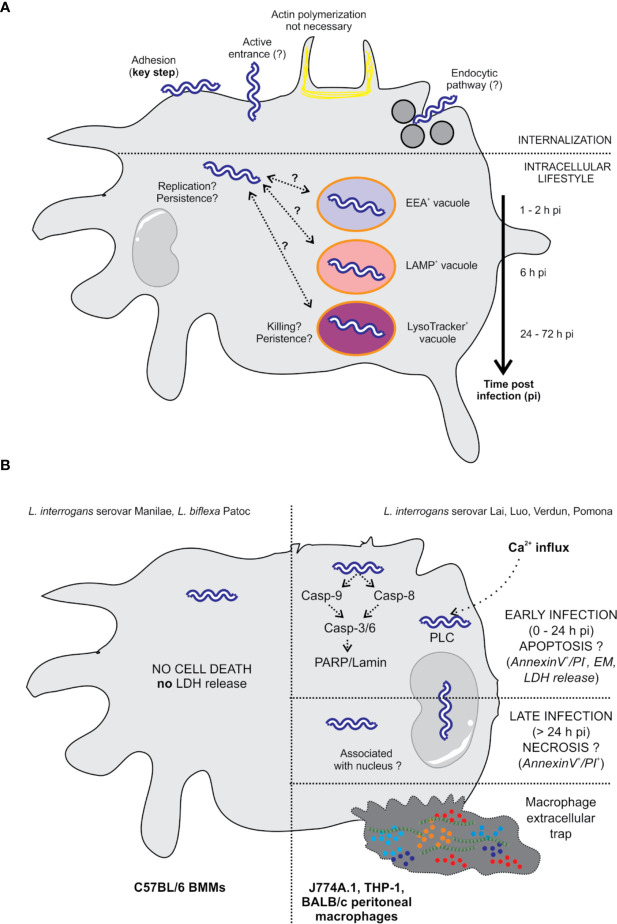
Figure 2.
Intracellular localization of L. interrogans and effect on cell death. (A) The internalization of leptospires is highly dependent on adhesion to the cell surface (25). The entrance mechanism is not clearly established since it does not seem to involve actin polymerization, suggesting an entrance mechanism other than phagocytosis (18, 25, 26). Viability seems to be essential since formalin-fixed bacteria do not enter the cells (18). The endocytic pathway could potentially be involved in the internalization of leptospires since inhibition of this pathway in macrophages drastically reduced the number of intracellular bacteria (18). Once inside the cells, leptospires are found free in the cytosol in human and murine macrophages. In addition, some authors describe that leptospires are found in EEA1-, LAMP-, and LysoTracker-positive compartments using colocalization (20, 24). Colocalization seems to be extended over time, which may suggest the arrest of potential phagosomes/autophagosomes at several stages. In human cells, leptospires appear to be replicative (20). However, in murine cells, the overall number of leptospires seems to be constant or diminished over time (20, 24), with the unique observation that live bacteria are still found at 72 hpi, suggesting their persistence. (B) L. interrogans serovar Manilae does not induce macrophage cell death upon infection of BMMs obtained from a C57BL/6 background (24). L. interrogans serovars Lai, Luo, and Verdun induce cell death by apoptosis/necrosis upon infection of J774A.1, THP-1, and BALB/c peritoneal macrophages (18, 21–23). However, not all the studies have completely correlating results, and some controversies are revealed in the literature. Additionally, some studies report that leptospires are associated with the nucleus (18, 23).

