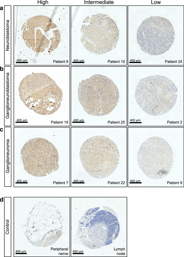
Figure 5.
Tissue microarray of different stages of NB shows a heterogeneous expression of anti-GD2 between patients. (a–d) Representative images of anti-GD2 immunohistochemical staining on a tissue microarray (TMA) containing tissue of 28 patients in duplicate. (a) Three representative images of differential GD2 labelling (high, intermediate and low) for neuroblastoma tissues. (b) Three representative images of differential labelling for ganglioneuroblastoma tissues and (c) three representative images for ganglioneuroma tissues. (d) Anti-GD2 stainings on control peripheral nerve (left) and lymph node tissue (right).

