Figure 1.
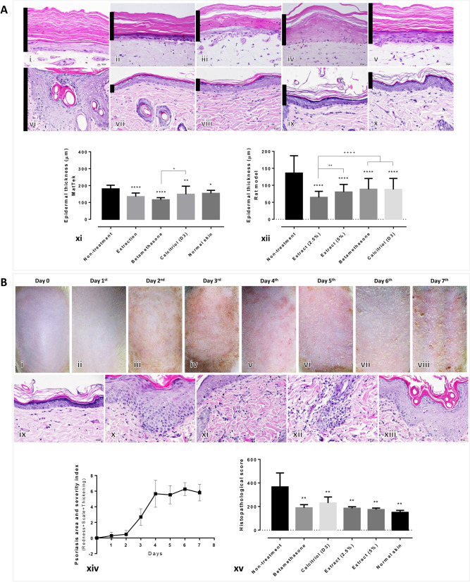
Histopathological evaluation of specimens from artificial psoriatic human skin and imiquimod-induced psoriatic rat skin: (A) Epidermal thickness at day 7 post-treatment in the artificial psoriatic (i–v) and rat psoriatic (xi, x) skins stained with hematoxylin and eosin (magnification, × 400). (i, vi) non-treatment; (ii, vii) extraction; (iii, viii) betamethasone; (iv, ix) calcitriol (D3); (v, x) normal skin groups. Bar graphs comparing the epidermal thickness in the artificial psoriatic (xi) and rat psoriatic (xii) skins. (B) Anatomical and histopathological appearances in imiquimod-induced psoriasis rats; gross skin lesion after induction with 62.5 mg of imiquimod from day 0 to day 7 (i–viii). Histopathological changes in rat psoriatic skin after 7 days of induction (ix–xiii). Ix, normal epidermis; x, acanthosis; xi, dermatitis; xii, folliculitis; xiii, hyperkeratosis. (xiv, xv) Line graph comparing the psoriatic area and severity index from day 0 to day 7 post-induction in each group (xiv). Bar graph comparing the histopathological scores among each treatment group (xv).

