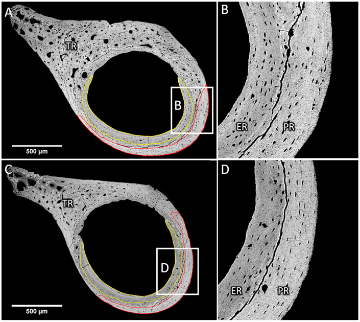
Fig. 2.
Femoral cross-sections of wild-type (A, B) and Brtl/+ (C, D) mice. Typical delimitation of endocortical (ER) and periosteal (PR) bone observed on a qBEI picture with high contrast at low (A, C) and high (B, D) magnification. The cortical thickness in Brtl/+ mice is smaller than in wild-type mice. ER and PR are separated by remnants of cartilage tissue resulting from the early phase of bone development. Noteworthy are the cracks due to sample preparation followed the delimitation. The third trochanter (TR) was excluded from the measurement since it exhibits a high porosity, high mineralization heterogeneity and numerous cartilage remnants.

