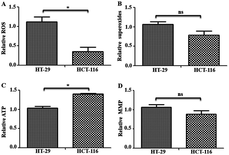
Figure 4.
Functional status of mitochondria in colorectal cancer cells. Cells (~1×10⁶) were used for mitochondrial functional analysis. (A) Total ROS levels were measured via staining with 2′,7′-dichlorodihydrofluorescein diacetate, and (B) mitochondrial superoxide levels were measured using MitoSOX™ Red staining followed by measurement on fluorescence plate reader. In both cases, cells were counterstained with Hoechst-33342 and used for normalization. (C) Total cellular ATP content was measured in cells with a luciferase-based ATP detection kit, and normalized with total protein levels. (D) MMP was measured after tetramethylrhodamine methyl ester perchlorate staining and normalization using Hoechs-33342 reading. *P<0.05. ns, not significant; ROS, reactive oxygen species; MMP, Mitochondrial Membrane Potential.

