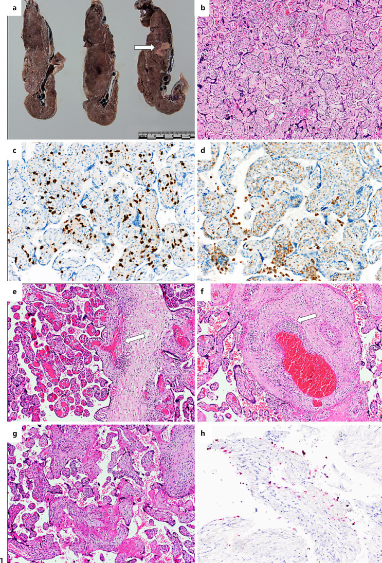
Fig. 1.
Findings of the placenta with manifest COVID-19. a Macroscopic image showing inhomogeneous and unusually condensed placental parenchyma and an area of infarction (arrow). b Chronic villitis and intervillositis (haematoxylin and eosin [H&E], 40×). c, d Characterisation of the inflammatory infiltrate consisting primarily of cytotoxic T-cells expressing CD8 (c) and fewer macrophages expressing CD68 (d) (immunohistochemistry, 200×). e, f Lymphohistiocytic villitis resulting in chorionic vasculitis and subsequent fresh (e) and already organizing thrombosis (f) (H&E, 100×). g Intervillous increase of fibrin as result of maternal malperfusion (H&E, 100×). h Presence of SARS-CoV-2 in decidual cells (red) (in situ hybridization for SARS-CoV-2, 200×).

