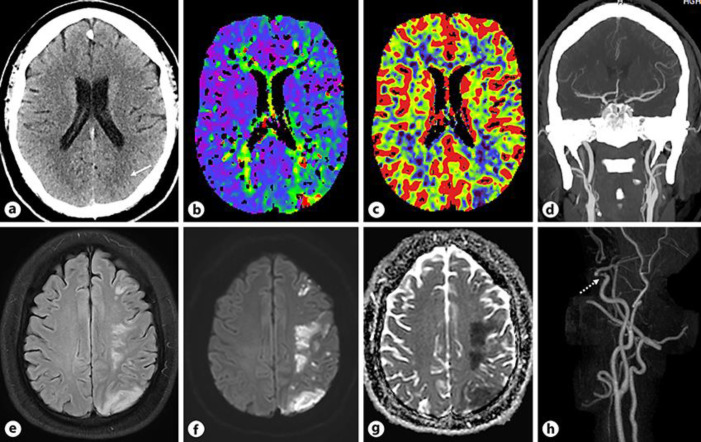
Fig. 2.
aAxial non-contrast CT section of the head shows early ischemic changes in the left posterior parietal lobe along the MCA-PCA watershed zone (white arrow). b, cAxial Tmaxand CBV perfusion maps demonstrate matched defect in the left posterior parietal lobe. dCoronal CTA reconstruction of the head reveals patent intracranial arteries. eAxial FLAIR image of the head demonstrates multiple patchy areas of increased signal in the left frontoparietal lobe along the watershed zone. f, gAxial DWI and ADC map show multiple acute infarcts in the left frontoparietal lobe. hPost contrast MRA 3D reconstruction of the neck and head reveals focal moderate to severe stenosis in the left distal cavernous ICA (dotted white arrow).

