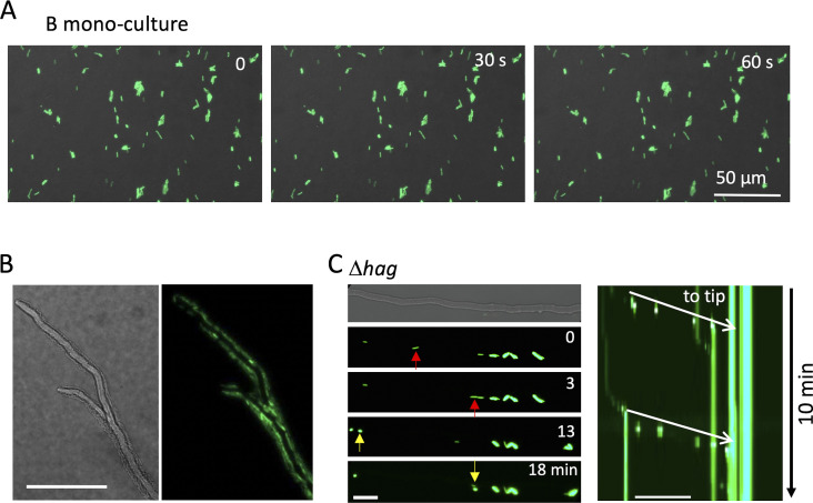
Figure S3. B. subtilis movement along hyphae by flagella.
(A) Time-lapse images of B. subtilis (green) monoculture for 60 s on the minimum agar media. Scale bar: 50 μm. (B) A. nidulans hyphae (DIC) surrounded by moving B. subtilis (green) from Video 4. (C) Image sequence of B. subtilis flagella mutant (Δhag) flow (arrows) along A. nidulans hyphae. Kymograph of the Δhag along the hyphae to the tip (white arrows) from Video 5. Total 10 min. Scale bar: 50 μm.

