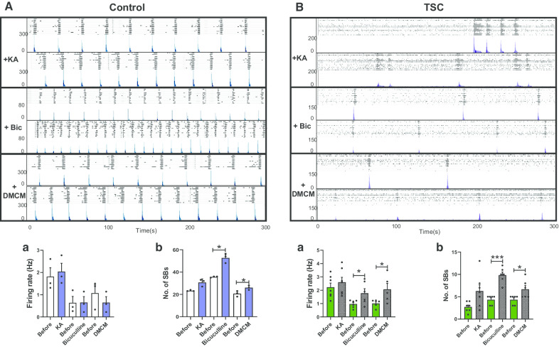
Fig. 2.
Pharmacological profiling of the network activity of TSC2 neurons. Typical synchronised burst patterns shown by raster plot (upper panel) and ASDR plots (lower patterns) for Control (A) and TSC2 (B) neurons, treated with 1 μM kainic acid (KA), 10 μM bicuculline (Bic) or 1 μM methyl-6,7-dimethoxy-4-ethyl-beta-carboline-3-carboxylate (DMCM). Plots (underneath) show mean ± SEM of a spike firing rate (Hz) and b number of synchronised bursts (SB) for all drugs. *p < 0.05, ***p < 0.001 following paired t-tests. Number of recorded wells = 3–10

