Fig. 1.

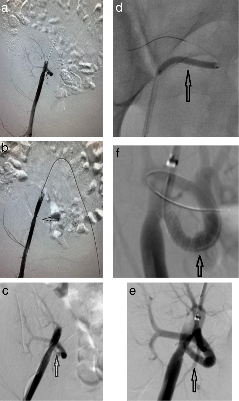
a and b show DSA images of the patient during the first endovascular treatment, demonstrating significant stenosis of 95% in the proximal transplant renal artery and transplant renal artery occlusion for repeated manipulations. c shows an angiographic image 25 days after the first operation, demonstrating spontaneous recanalization of the transplant renal artery. d shows a DSA image of the patient during the second endovascular treatment, demonstrating that a 3 mm × 30 mm balloon (Boston ultra-soft SV) was used to dilate the stenotic segment. e and f show DSA images of the patient, demonstrating that a 4 mm × 23 mm stent (Firehawk Rapamycin Target Eluting Coronary Stent) was implanted successfully in the stenotic segment and that the transplant renal artery was well reconstructed
