
Figure 2: Platelet lamellipodial ring tension is driven by a thin ring of polymerizing actin.
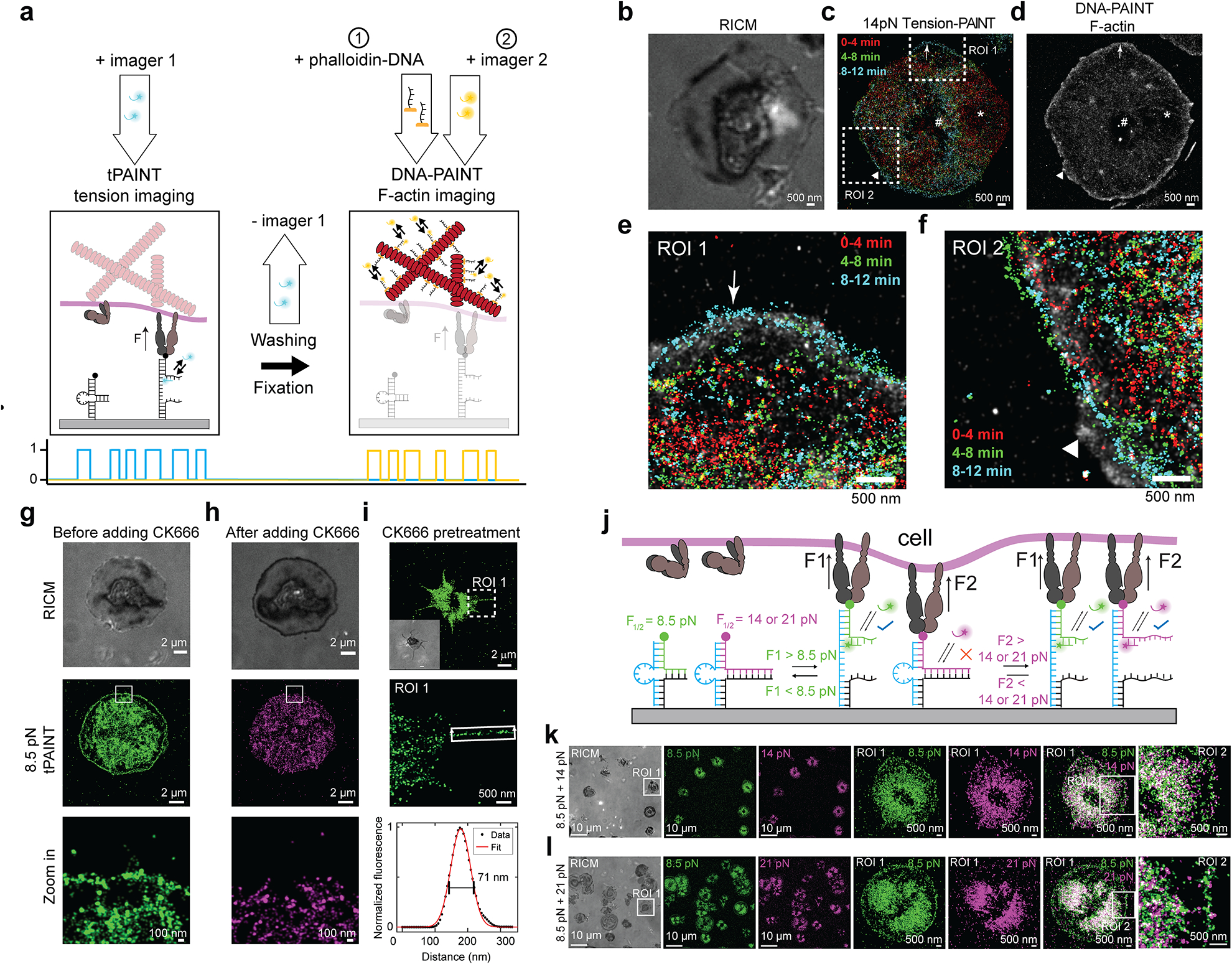
(a) Platelet 14 pN integrin tension was imaged before fixation, staining, and Exchange-PAINT to super-resolve the platelet actin cytoskeleton. (b) RICM, (c) time-resolved 14pN tension-PAINT, and (d) F-actin stained with DNA-tagged phalloidin. (e-f) Zoom in of tension at the lamellipodial edge reveals close association between tension and actin (arrow). In some regions, actin polymerization extends beyond tension (triangle). Note that an area of depleted actin (*) corresponds to an area where tension receded after 4 min. The # indicates a fiducial marker. Actin-tension data is representative of 7 platelets from n = 2 independent experiments. Note that tension-actin localizations use higher contrast for tension than F-actin because F-actin localization density is higher than tension localization density. (g-h) Treatment with the Arp2/3 inhibitor CK666 removes platelet lamellipodial edge tension (2 platelets) or prevents lamellipodial edge tension (7 platelets) (9 platelets total from n = 2 independent experiments) (i) Platelets pretreated with 50 μM CK666 spread slowly, utilize filopodia to engage with the cRGD substrate, and do not exhibit lamellipodial edge tension (18 platelets from n = 3 independent experiments). (j) Force-multiplexed tPAINT is possible by coating surfaces in two probes with F1/2 of 8.5 and 14 or 21pN, (k-l) and reveals that lamellipodial edge tension exceeds 14pN, but does not frequently exceed 21pN. Integrin tension in the center of platelets exceeds 21pN. The data shown in Inhibition studies and force-multiplexed tPAINT are representative of n = 3 independent experiments.