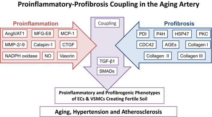
Figure 1.

Diagram depicting the arterial proinflammatory‐profibrosis coupling events. AGEs, advanced glycosylation end‐products; AngII, angiotensin II; AT1, Ang II type 1 receptor; CDC42, cell division control protein 42 homolog; CTGF, connective tissue growth factor; ECs, endothelial cells; HSP47, heat shock protein 47; MCP‐1, monocyte chemoattractant protein‐1; MFG‐E8, milk fat globule epidermal growth factor‐8; MMPs, matrix metalloproteinases; NADPH oxidase, nicotinamide adenine dinucleotide phosphate oxidase; NO, nitric oxide; P4H, prolyl‐4‐hydroxylase; PDI, protein‐disulfide isomerase; PKC, protein kinase C; SMADs, homologies to the Caenorhabditis elegans SMA (“small” worm phenotype) and Drosophila MAD (“Mothers Against Decapentaplegic”) family of proteins; TGF‐β1, transforming growth factor‐beta 1; VSMCs, vascular smooth muscle cells.
