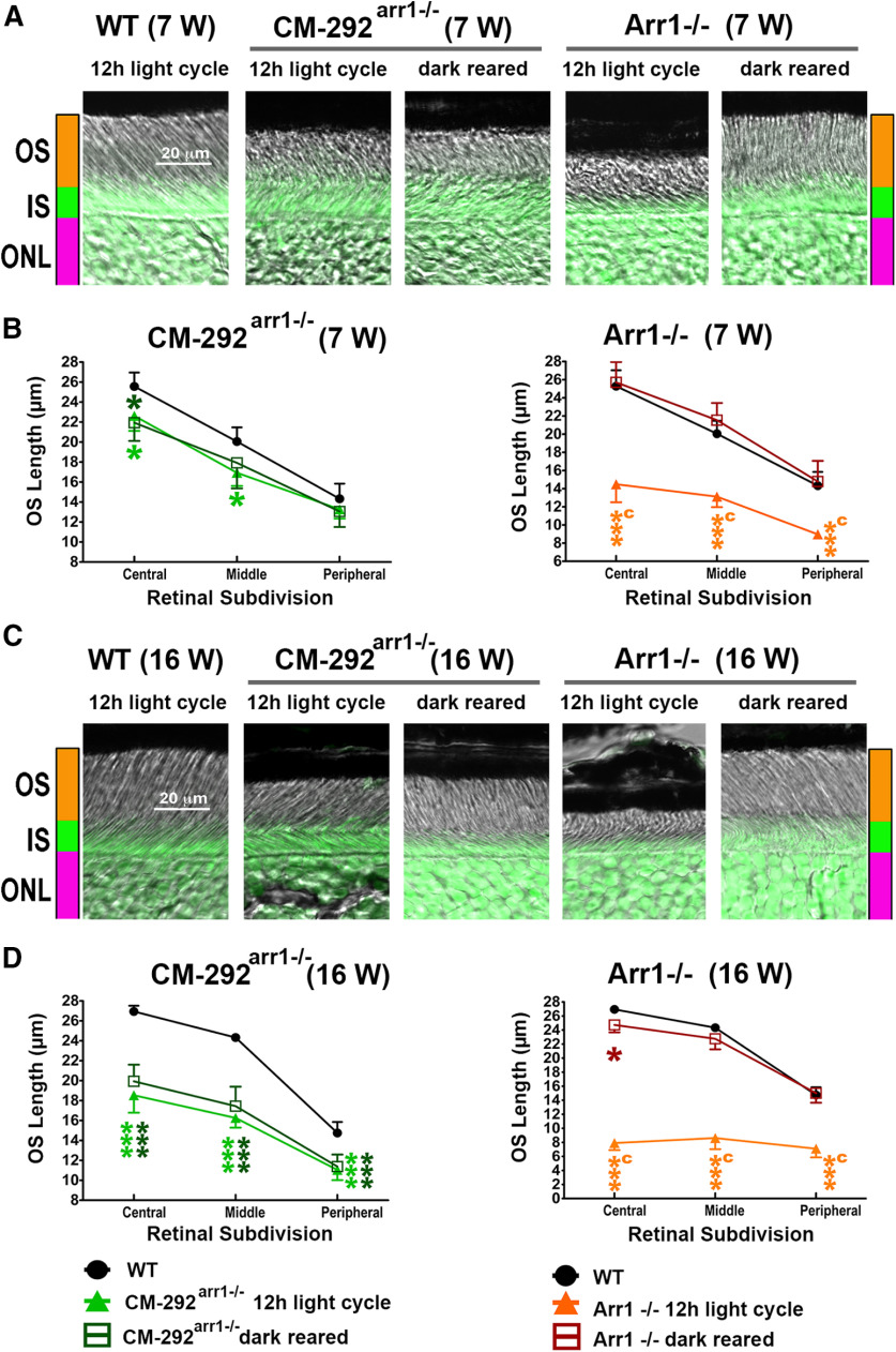
Figure 11.
The damage caused by supraphysiological expression of CM is light independent. A, B, Combined DIC and green fluorescent Nissl images of the retina sections of 7-week-old (A) and 16-week-old (B) mice of indicated genotypes, enlarged to show the OS more clearly. The positions of OSs, ISs, and ONL are shown on the left. C, D, The length of the OS measured in the central, middle, and peripheral retina were the average of inferior and superior hemispheres. Mean ± SEM values from five animals per genotype are shown. The length of OS was compared separately for each age by two-way repeated-measures ANOVA with retinal subdivision as the within-group factor and genotype as the between-group factor followed by Bonferroni post hoc test for each retinal subdivision. The CM-292arr1−/− mice were significantly different from WT mice regardless of light exposure at both ages across retinal subdivisions (p < 0.01 at 7 weeks; p < 0.0001 at 16 weeks). In contrast, Arr1−/− mice kept in a normal light cycle significantly differed from WT mice at both ages (p < 0.0001). The dark-reared Arr1−/− mice were no different from WT mice but were significantly different from light-exposed mice at both ages (p < 0.0001). Statistical significance of the differences is shown, as follows (color coded): *p < 0.05; ***p < 0.001 compared with WT mice; a p < 0.05, c p < 0.001 compared with Arr1−/− dark mice. Note that dark rearing preserves the OS in Arr1−/− mice, whereas the damage in CM-292Arr−/− animals is not affected by light.

