
Figure 5.
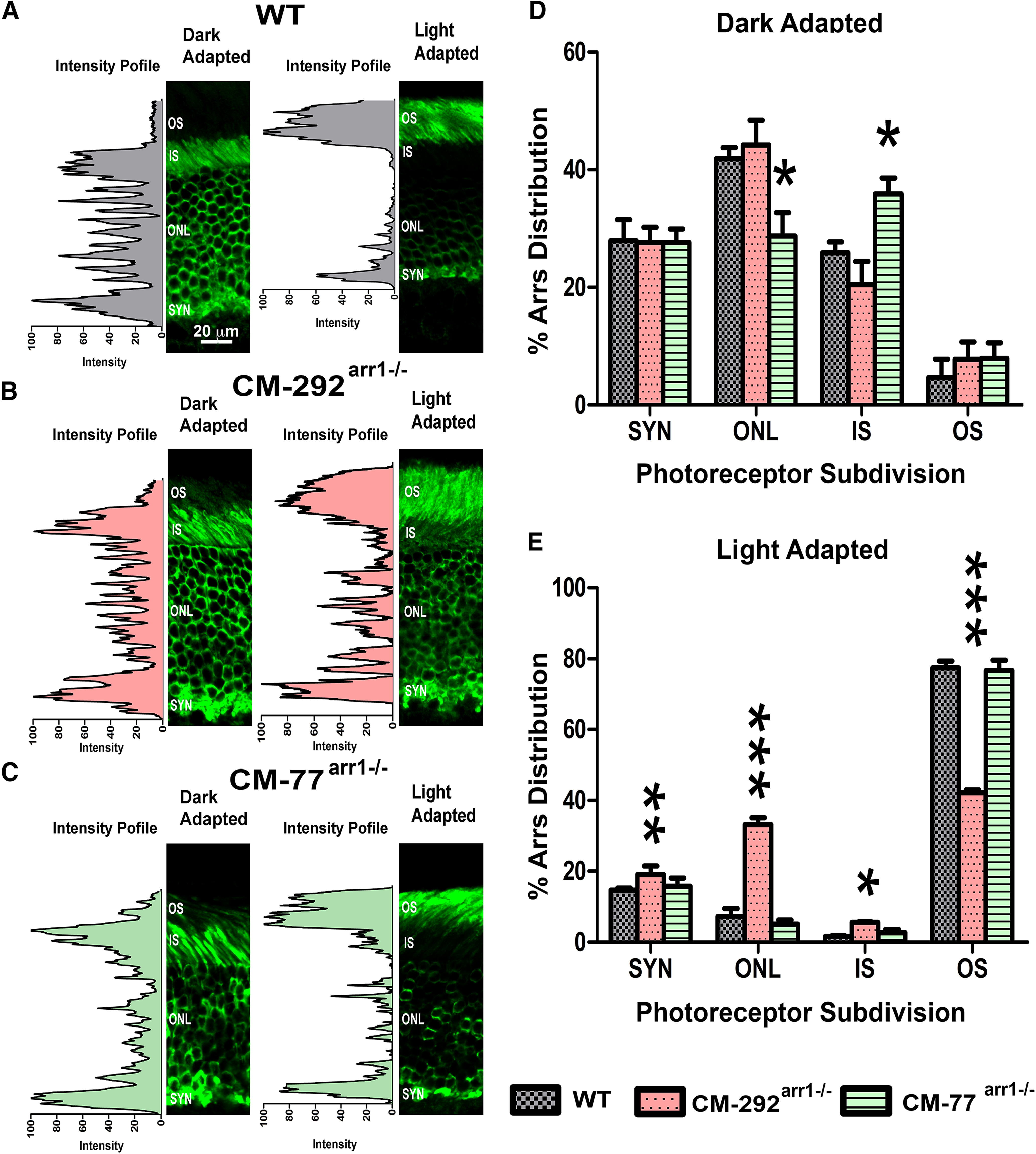
Arrestin-1 localization in the dark- and light-adapted rod photoreceptors. A–C, Mice with the indicated genotypes (A–C) were dark adapted overnight (DARK) or exposed to light (2700 lux for 1 h; LIGHT). Eyes were fixed and processed for Arr1 immunohistochemistry using C10C10 anti-arrestin-1 antibody (Knospe et al., 1988). The positions of the OSs, ISs, ONL, and synaptic layer (SYN) are indicated. D, E, The proportion of Arr1 localized in the OSs of dark-adapted (D) and light-adapted (E) mice was quantified by the intensity of Arr1 immunostaining (green) in three images per animal from three animals per genotype. Mean ± SD values are shown. The two-way ANOVA with genotype and rod compartment as main factors yielded no significant effect of genotype (p = 0.99) but a highly significant genotype × compartment interaction (p < 0.0001). *p < 0.05; **p < 0.01; ***p < 0.001, compared with the corresponding compartment in WT mice, as detected by independent multiple comparison of means with Dunnett's multiple-comparison tests.