Figure 4.

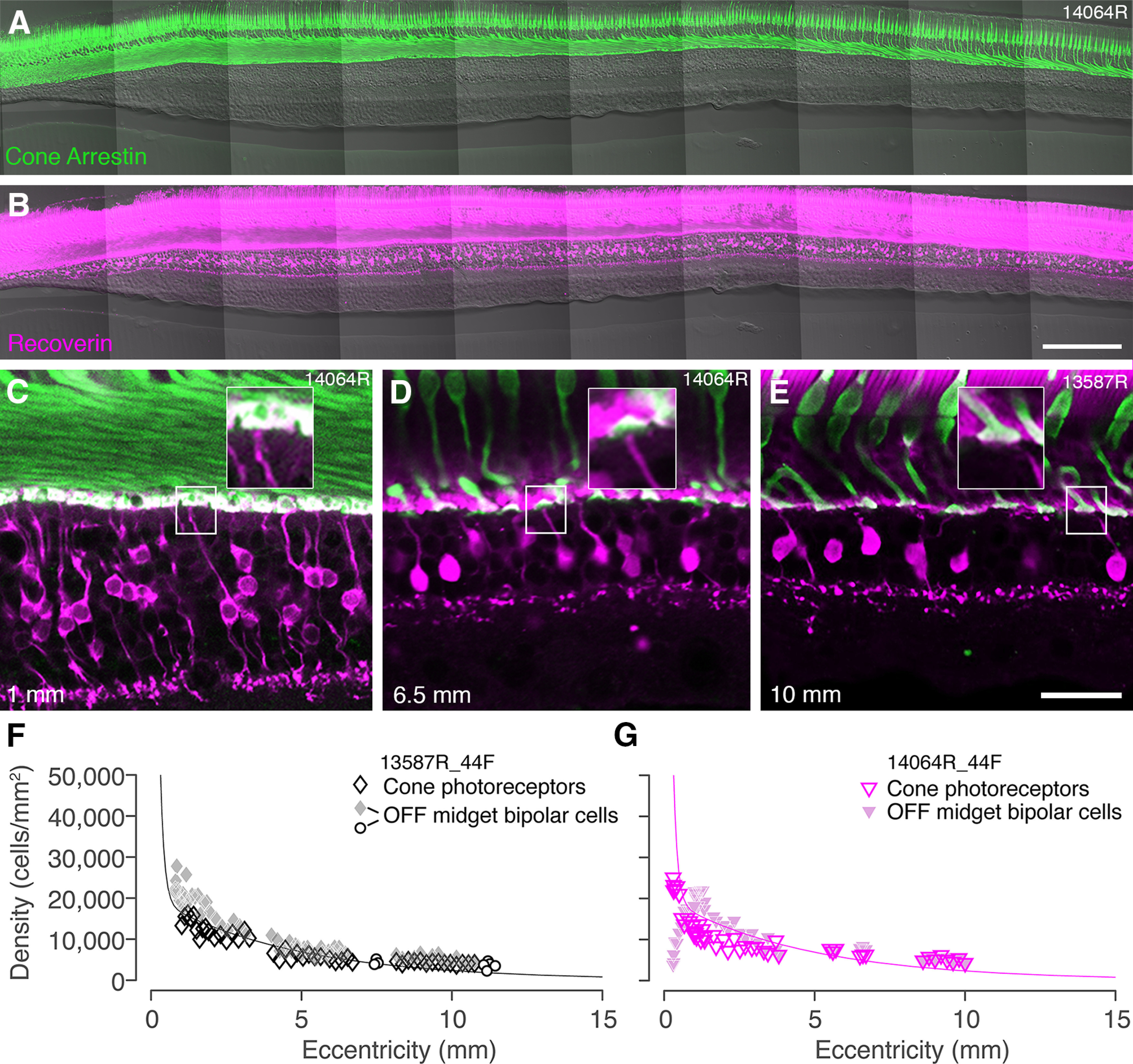
Convergence of cones to midget bipolar cells. A, B, Confocal images of a vertical vibratome section through temporal retina 300 µm superior to the fovea of preparation #14064 stained with antibodies against cone arrestin (A) and recoverin (B). C–E, Confocal images from sections double labeled for cone arrestin and recoverin to illustrate connectivity between cone photoreceptors and OFF-midget bipolar cells at various eccentricities. Insets in each image show that single-headed flat midget bipolar cells can be identified up to at least 10-mm eccentricity. Eccentricity is indicated in the lower left corner. F, G, Cell density is plotted against eccentricity for cone photoreceptors and flat midget bipolar cells in preparation #13587 (F) and #14064 (G). Open circle symbols in panel F show values obtained from whole mounts, as described in the text. The curves represent the average density of cone photoreceptors across all preparations. In both preparations, the ratio of cones to OFF-midget bipolar cells is 1:1 in peripheral retina. Scale bars: 200 µm (B, applies to A, B) and 50 µm (E, applies to C–E).
