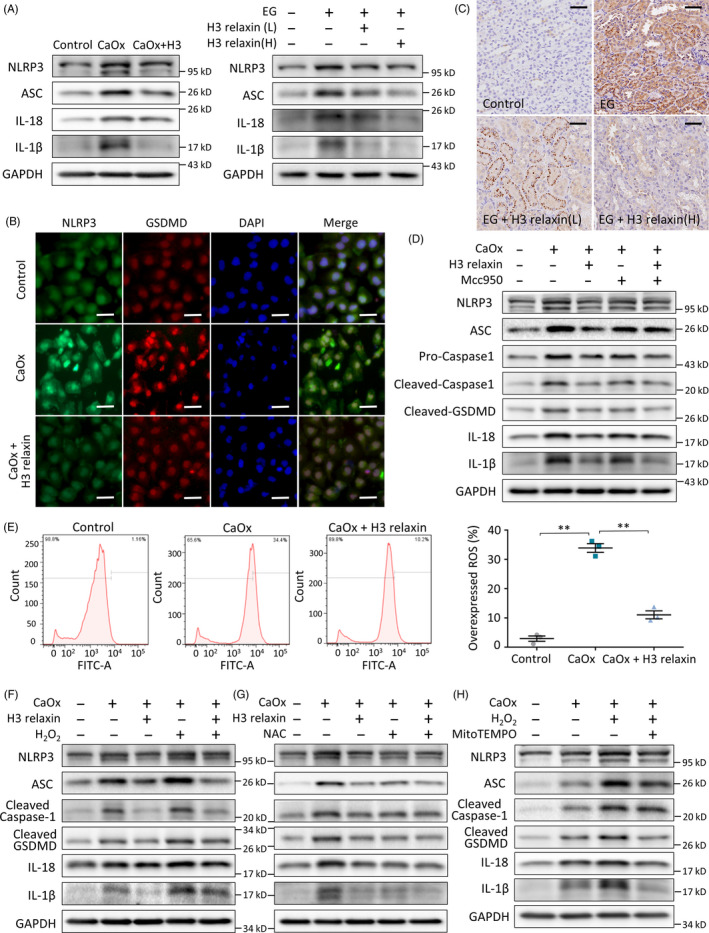
FIGURE 3.

H3 relaxin inhibits CaOx crystal‐induced pyroptosis by preventing the reactive oxygen species (ROS)‐mediated NLRP3 inflammasome activation. A, Western blot analysis of related proteins: NLRP3, ASC, IL‐18 and IL‐1β in the treated tubular epithelial cells (TECs) or rat kidney samples (14 d), in the absence or presence of H3 relaxin. B, The protein expression of NLRP3 and cleaved GSDMD was detected using immunofluorescence assays in the treated TECs in groups. Scale bars: 100 μm. C, The protein expression of NLRP3 was detected using immunohistochemical assays in the treated rat kidneys (14 d), with or without H3 relaxin treated. Scale bars: 50 μm. D, Western blot analysis of inflammation‐related proteins: NLRP3, ASC, cleaved caspase‐1, cleaved GSDMD, IL‐18 and IL‐1β in the treated TECs, in the absence or presence of H3 relaxin and Mcc950. n = 3 per group, representative images are shown. E, ROS generation of the treated TECs was detected by DCFH‐DA probe using flow cytometric analysis. n = 3, data expressed as means ± SEM, **P < .01. F, Western blot analysis of inflammatory pyroptosis‐related proteins in the treated TECs, in the absence or presence of H3 relaxin and H2O2. G, Western blot analysis of inflammatory pyroptosis‐related proteins in the treated TECs, in the absence or presence of H3 relaxin and NAC. H, Western blot analysis of inflammatory pyroptosis‐related proteins in the treated TECs, in the absence or presence of H2O2 and MitoTEMPO. n = 3 per group, representative images are shown
