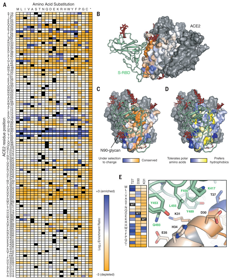
Fig. 1. Sequence preferences of ACE2 residues for high binding to the RBD of SARS-CoV-2 S.
(A) Log2 enrichment ratios from the nCoV-S-High sorts are plotted from depleted or deleterious (orange) to enriched (dark blue). ACE2 primary structure is shown on the vertical axis, amino acid substitutions are indicated on the horizontal axis. Wild-type amino acids are in black. Asterisk (*) denotes stop codon. (B) Conservation scores are mapped to the structure (Protein Data Bank 6M17) of RBD (green ribbon)–bound protease domain (surface), oriented with the substrate-binding cavity facing the reader. Residues conserved for RBD binding are shown in orange; mutationally tolerant residues are in pale colors; residues that are hot spots for enriched mutations are in blue; and residues maintained as wild type in the ACE2 library are in gray. Glycans are depicted as dark red sticks. (C) Viewed looking down on to the RBD interaction surface. (D) Average hydrophobicity-weighted enrichment ratios are mapped to the structure, with residues tolerant of polar substitutions in blue and residues that prefer hydrophobics in yellow. (E) A magnified view of the ACE2–RBD interface [colored as in (B) and (C)]. Heat-map plots log2 enrichment ratios from the nCoV-S-High sort. Abbreviations for the amino acid residues are as follows: A, Ala; C, Cys; D, Asp; E, Glu; F, Phe; G, Gly; H, His; I, Ile; K, Lys; L, Leu; M, Met; N, Asn; P, Pro; Q, Gln; R, Arg; S, Ser; T, Thr; V, Val; W, Trp; and Y, Tyr.

