Figure 1:

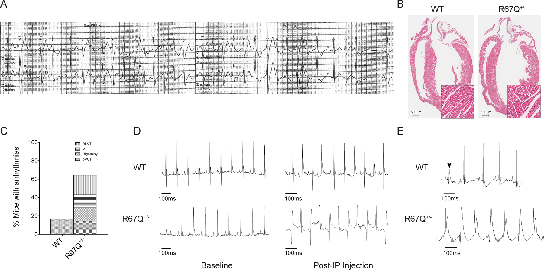
R67Q+/− mice develop paroxysmal bi-directional VT following administration of caffeine and epinephrine. A. Tracing from a Holter monitor of the patient with R67Q mutation showing BiVT and PMVT. B. Hematoxylin and Eosin staining of whole heart from WT and R67Q+/−. Gross histology revealed no significant difference between WT and R67Q+/− in heart structure and size. Scale bar is 500μm. Insert scale is 50μm C. Quantification of arrhythmic events observed in WT and R67Q+/− during ECG analysis. D. ECG recorded from anesthetized WT (top panel) and R67Q+/− (bottom panel) mice at baseline for 5 minutes. Normal sinus rhythm was observed in both groups. Epinephrine (2mg/kg) and caffeine (120mg/kg) were administered by IP injection and ECG recorded for up to 30 minutes. WT showed faster sinus rhythm (top right panel), whereas R67Q+/− mice developed paroxysm of bi-directional ventricular tachycardia (bottom right panel taken at 10 minutes post-injection). E. Upper trace shows a representative PVC observed in WT mice following injection of caffeine and epinephrine. PVC is highlight by the arrow. Lower trace shows representative polymorphic ventricular tachycardia observed in R67Q+/− following injection. N=8 animals per group. Bi-VT – bi-directional ventricular tachycardia; VT – ventricular tachycardia; pVCs – premature ventricular contractions; IP – intraperitoneal injection.
