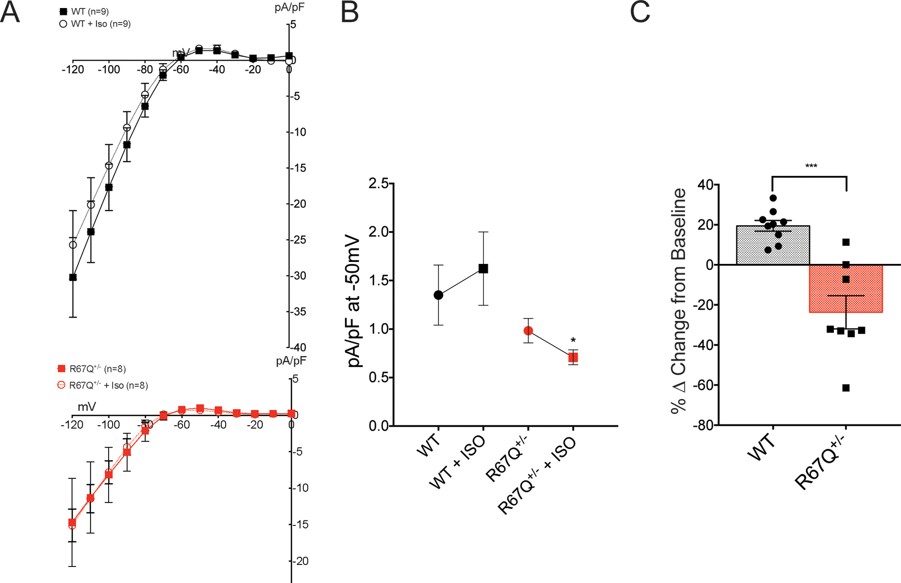
Figure 3:

IK1 fails to increase following isoproterenol. A. Baseline current voltage relationship for WT (black squares, N=5, n=9), R67Q+/− (red squares, N=5, n=8). Following isoproterenol (ISO) perfusion for 5 minutes, WT outward current (black open circles, N=5, n=9) increased, however R67Q+/− outward current (red open circles, N=5, n=8) decreased in response to adrenergic stress. Currents shown are calculated following barium subtraction. Barium is perfused for 2 minutes following final ISO measurement. B. Absolute current values at −50mV following ISO treatment: R67Q+/− outward IK1 (0.71 ± 0.08 pA/pF) is significantly decreased compared to WT (1.62 ± 0.38 pA/pF) following isoproterenol treatment (p<0.05). C. Peak outward current was determined at −50 mV and the percentage change for each cell from baseline was calculated. The delta from baseline showed a 19.5 ± 2.7% increase in outward current at −50mV in WT cells (black, N=5, n=9), whereas R67Q+/− cells (red, N=5, n=8) showed a 23.7 ± 8.3% decrease in outward current at −50 mV. *** p<0.001. Students t-test was used to determine significance difference between groups (3B), or Two-way repeated measures ANOVA with post-hoc Bonferroni correction was used (3C).
