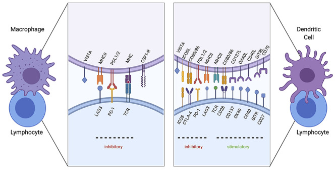
Figure 1.

Immune checkpoint signaling under investigation. Validated checkpoint marker interactions between macrophages (left) and dendritic cells (right) with lymphocytes are depicted. TCR interacting with MHC II serves as the first stimulation signal. Macrophages can play an immunosuppressive role by upregulating the surface expression of checkpoint proteins PD-L1 and ligand 2 (PD-L2), which interacts with Programmed cell death proten 1 (PD-1) on lymphocytes. Furthermore, lymphocyte-activation gene 3 (LAG3) will bind to MHC II molecules and reduce monocyte differentiation. Colony-stimulating factor 1 receptor (CSF1R) also functions to differentiate monocytes to macrophages. V-domain immunoglobulin suppressor of T cell activation (VISTA) functions to dampen T-cell activation. A secondary signal between dendritic cells and lymphocytes will determine the stimulatory or inhibitory response. Stimulatory checkpoint pairs include CD28 and CD80/86, CD137/CD137 ligand, OX40/OX40 ligand, CD40/CD40 ligand, CD27/CD70 and glucocorticoid-induced tumor necrosis factor receptor (GITR)/GITR ligand. Inhibitory checkpoint pairs include inducible T cell co-stimulator (ICOS) with ICOS ligand, cytotoxic T-lymphocyte-associated antigen 4 (CTLA-4)/CD80 or 86, PD-1/PDL1/2, LAG3/MHC II and VISTA (Created with BioRender.com).
