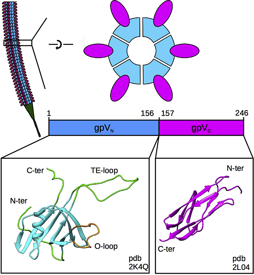
Fig. 1. Organization of the lambda tail.
Top left, representation of the tail tube showing identical disks of the gpV tail tube protein stacked along the length of the tail and the tail tip in green. Top right, top, and side views of a single hexameric disk of the tail tube protein showing the organization of the N-terminal gpVN (blue) and C-terminal gpVC (magenta) domains. Bottom, the monomeric NMR structures of the gpVN domain (PDB ID: 2K4Q), including the rigid (blue), flexible (green) and O-loop (tan) regions, and the Ig-like gpVC domain (PDB ID: 2L04).

