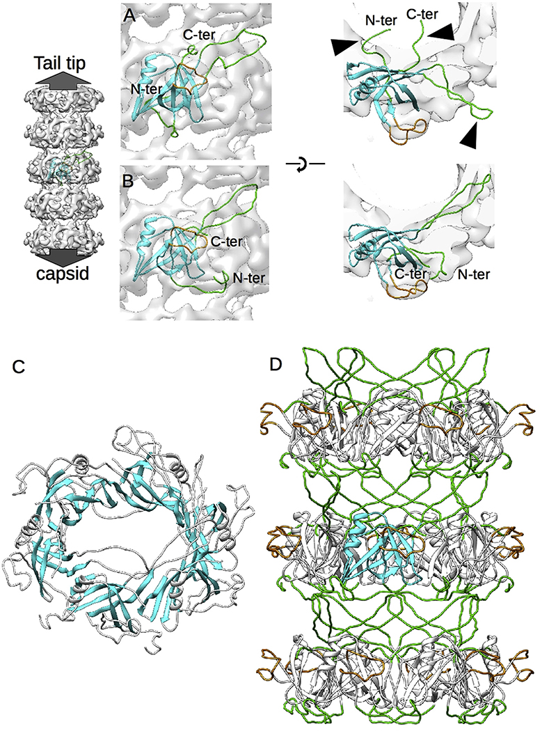
Fig. 3. Modeling of the N-terminal gpVN domain.
A. Rigid body fitting of the gpVN domain model (PDB Id: 2K4Q) into the free tail density map viewed from the side of the tube (left) and the top (right). One representative NMR model of the 20 available has been chosen. B. Same views as (A) after refinement of the gpVN structure by flexible fitting. C. Formation of an intradisk beta barrel-like structure mediated by the rigid beta strands of the gpVN domain. D. Final model of the inner tail tube structure, showing that the interdisk contacts are mediated by the flexible loops (green) identified in the NMR model of gpVN.

