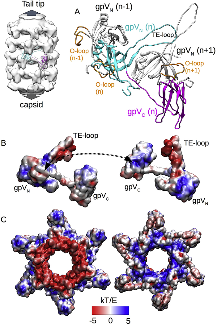
Fig. 5. Electrostatic forces stabilize the tail tube structure.
A. Structural model of the entire gpV including both N-terminal gpVN and C-terminal gpVC domains as well as gpVN domains from two adjacent gpV subunits. B. Electrostatic pattern of the gpV subunit viewed from the outside (left) and inside (right) of the tail tube. A dashed double arrow shows the contact area of the O-loop and the Ig-like domain of an adjacent subunit. C. Electrostatic pattern of a tail tube disk viewed from the tail-tip end (left) or the capsid end (right) of the tail tube.

