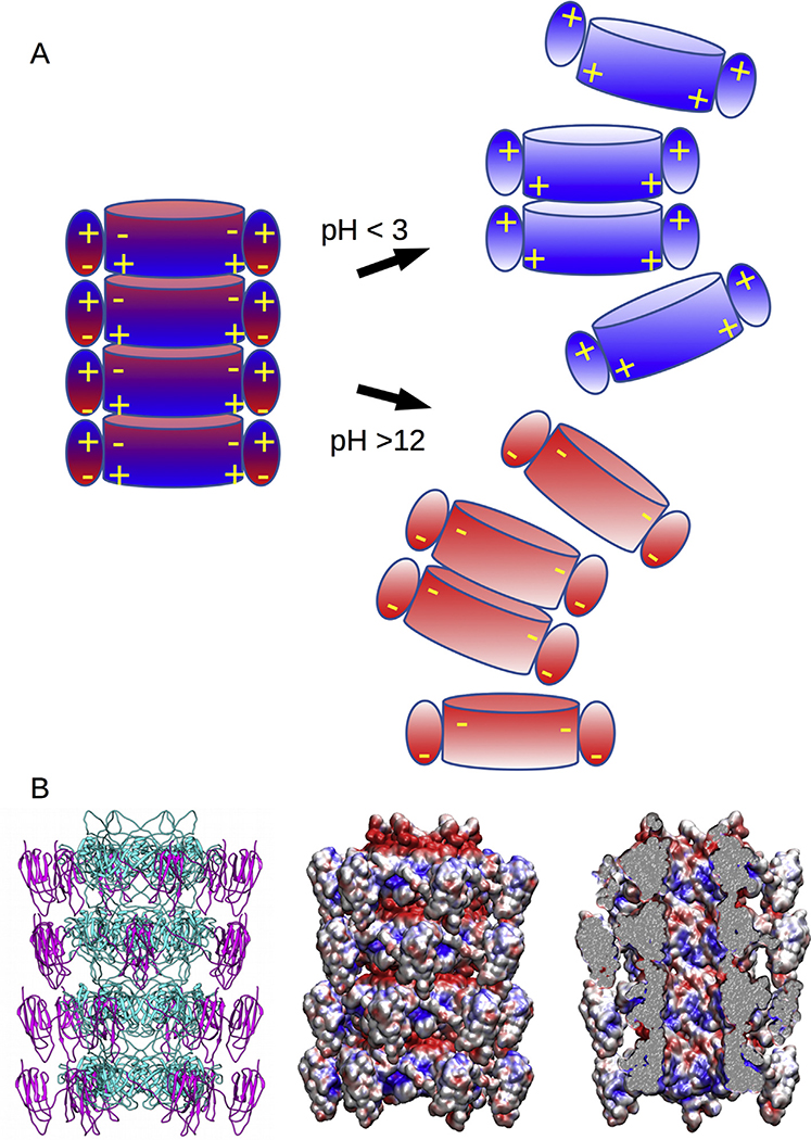
Fig. 6. Tail tube structure and disassembly model.
A. Disassembly model of the tail tube because of the masking of charges on either the inner tube at low pH, or the Ig-like domain at high pH, and the subsequent charge repulsion between disks. B. Structural model (right) of the tail tube with the electrostatic patterns shown viewed from the outside (middle) and inside (right) of the tail tube showing that its lumen surface is composed of a mix of negative and positive charges.

