Fig. 1.
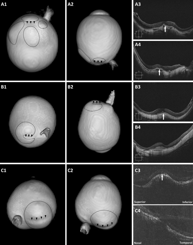
Eyes with a DSM on OCT and a macular inward convexity on 3D MRI. A1 and A2. Posterior view and side view of 3D MRI of the eye. One macular inward convexity (arrowheads) and four distinct staphylomas (circles) around are observed; (A3 and A4) both vertical and horizontal scans show a convex macula (arrows). B1 and B2. Posterior view and side view of 3D MRI of the eye. A band-shaped inward convexity (arrowheads) and a kidney-shaped staphyloma (circle) nearby are observed; (B3) vertical scan shows a clearly convex macula (arrow); (B4) horizontal scan shows a flat macula. C1 and C2. Posterior view and side view of 3D MRI of the eye. The staphyloma (circle) was horizontally split into two parts by the band-shaped inward convexity (arrowheads); (C3) vertical scan shows a clearly convex macula (arrow); (C4) horizontal scan shows a slightly convex macula.

