Fig. 2.
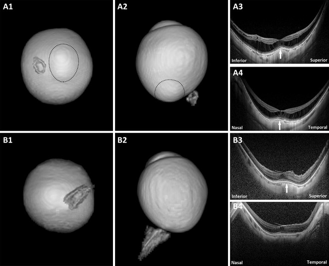
Eyes with a DSM on OCT but with no macular inward convexity on 3D MRI. A1 and A2. Posterior view and side view of 3D MRI of the eye. A staphyloma (circle) is observed; (A3 and A4) both vertical and horizontal scans show a convex macula (arrows) and obvious macular retinoschisis. No obvious dome-shaped convexity of the vitreoretinal interface is observed. B1 and B2. Posterior view and side view of 3D MRI of the eye. No protrusion or inward convexity is observed; (B3) vertical scan shows a clearly convex macula (arrow) and obvious macular pucker and a small foveal cystoid space. No obvious dome-shaped convexity of the vitreoretinal interface is observed; (B4) horizontal scan shows a flat macula and obvious macular pucker and a small foveal cystoid space.

