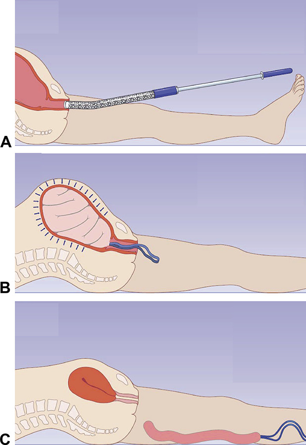
Fig. 2. Placement and removal of study device. The applicator is inserted through the vagina into the lower uterine segment. A. The plunger is depressed, ejecting the mini-sponge pouch into the uterus. B. The applicator is gently removed from the uterus, and the removal strand is left in the vaginal canal. Once inside the uterus, the mini-sponges absorb blood and pack the uterus. C. When the patient is clinically stable and it is deemed safe to remove the dressing, the pouch is removed by applying gentle, steady traction to the removal strand. Images courtesy of OBSTETRX. Used with permission.

Rodriguez. Mini Sponge Device for Uterine Tamponade. Obstet Gynecol 2020.
