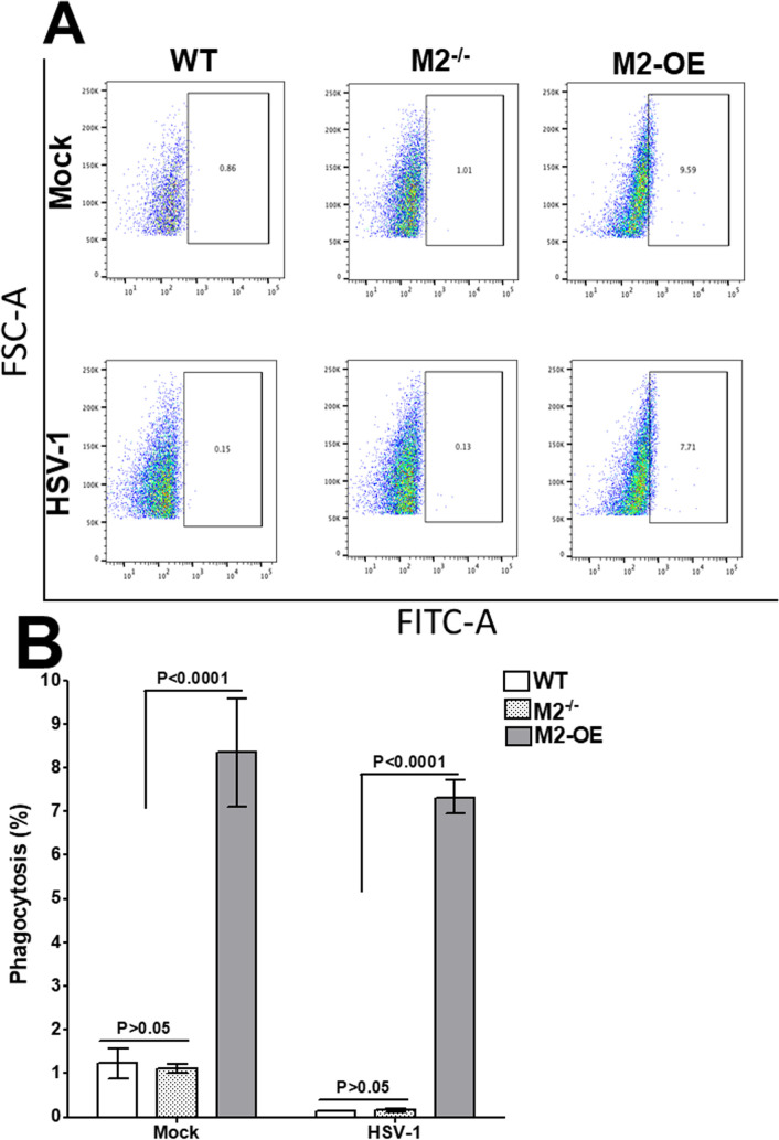
Fig 3. Phagocytosis assay on in vitro derived bone marrow-derived (BM) macrophages.
Cells from BM-derived WT, M2-/-, and M2-OE mice were cultured, differentiated into macrophages, and cultured in 24-well plates as described in Materials and Methods. After overnight culture, adhered cells were infected with HSV-1 and incubated with latex beads-rabbit IgG-FITC complex. Cells were stained with F4/80 AF 564 antibody and subjected to flow cytometry analysis. Panel A) representative plots of WT, M2-/-, and M2-OE macrophages; and B) Percentage phagocytosis plots from three separate experiments.

