
FIGURE 2.
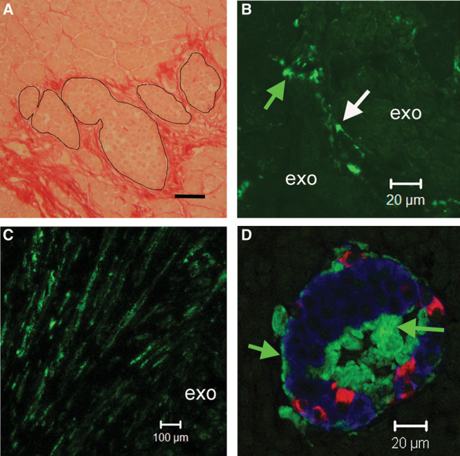
A, Explant stained (red) with Sirius red for collagen. Fibrotic tissue surrounded islets (outlined) and accumulated in interlobular and intralobular spaces; scale bar 50 µm. B, Cells (possibly pericytes) labeled for SMA (arrows) were present between exocrine lobules. C, SMA positive cells in interlobular fibrotic tracts between exocrine lobules (exo). D, Islet labeled for insulin (blue) glucagon (red) and islet amyloid (green). Islet amyloid was localized adjacent to islet capillaries at the islet periphery and in the center of the islet (green arrows). SMA, smooth muscle actin.