FIGURE 6:

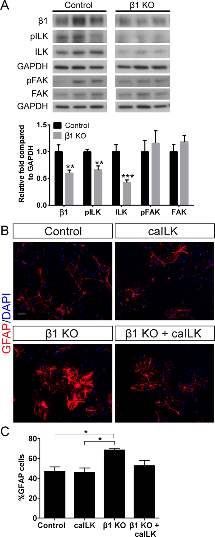
β1-integrin regulates astrocyte differentiation through ILK. A: Western blot of DG tissue after β1-integrin knockout as in Fig. 2A. β1-integrin ablation decreases levels of pILK and total ILK but does not change levels of pFAK or total FAK. GAPDH was the loading control and values are expressed as relative fold compared to control. B: Hippocampal NSC cultures from female β1+/+ ROSA:YFP or β1+/+; ROSA:YFP mice were infected with adenovirus expressing Cre (AV-Cre) to generate control and β1KO neurospheres. Lentivirus expressing constitutively active ILK (LV-caILK) was used to increase ILK signaling. Immunostaining for GFAP revealed that β1-integrin knockout increases astrocyte differentiation and that addition of caILK to β1 KO cultures rescues the increase in astrocyte differentiation. C: Quantification of GFAP+ cells shows that β1-integrin ablation increases the percentage of infected cells that differentiate into GFAP+ astrocytes compared to control, while addition of caILK to β1 KO cultures reduces astrocyte differentiation back towards control levels. Scale bar 50 µm. Data are means ± SEM. Unpaired Student’s t test: **P < 0.01, ***P < 0.001. One-way ANOVA with Tukey’s post hoc *P < 0.05. [Color figure can be viewed in the online issue, which is available at wileyonlinelibrary.com.]
