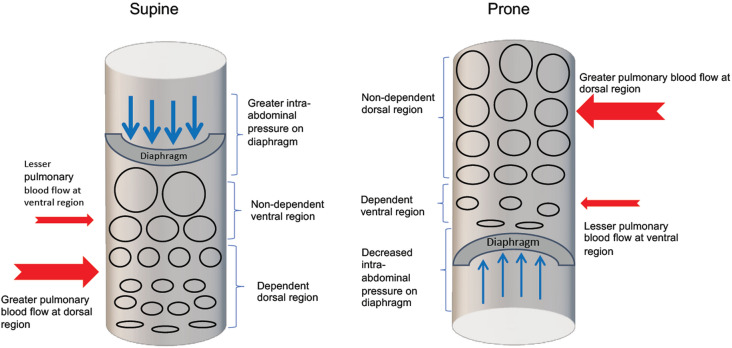
Fig. 1.
Schematic showing the changes in ventilation and perfusion in supine and prone positions. In the supine position, alveoli at the dependent dorsal region are collapsed (flattened ovals) resulting in decreased ventilation due to the compressive forces exerted by the ventral region lung tissues as well as the increased (thicker blue arrows) intra-abdominal pressure transmitted to the diaphragm. Greater pulmonary blood flow (thicker red arrow) and decreased ventilation at the dorsal region led to greater ventilation/perfusion mismatch. In the prone position, without the weight of the compressive forces of the ventral region and decreased intra-abdominal pressure (thinner blue arrows), alveoli at the now non-dependent dorsal region are recruited (bigger circles) and coupled with greater pulmonary blood flow (thicker red arrow) at the dorsal region, there is now better ventilation/perfusion matching thereby resulting in better oxygenation

