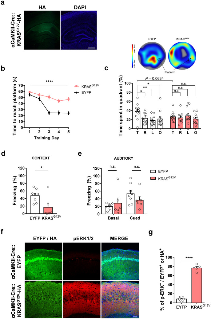
Figure 3.
Ectopic expression of KRASG12V in excitatory neurons impairs spatial memory and induces ERK activation. (a) HA staining of KRASG12V-expressing hippocampal slices from αCaMKII-Cre mice. 4′,6-diamidino-2-phenylindole (DAPI) staining was used to identify nuclei. Scale bars, 200 μm. (b) Learning is significantly slower in KRASG12V expressing mice compared to EYFP controls. αCaMKII-Cre::EYFP, n = 12; αCaMKII-Cre::KRASG12V, n = 11; Two-way repeated measures ANOVA, F1, 21 = 69.63, ****P < 0.001 (c) Lower panel, time spent in each quadrant during the probe test. αCaMKII-Cre::EYFP mice selectively searched for the platform in the target quadrant, while KRASG12V did not. αCaMKII-Cre::EYFP, n = 12, One-Way ANOVA, followed by a Dunnett’s post hoc test, *P = 0.0292, **P = 0.0030, *P = 0.0156; αCaMKII-Cre::KRASG12V, n = 11, One-way ANOVA, followed by Dunnett’s post hoc test, P = 0.9510, P = 0.9743, P = 0.7006. αCaMKII-Cre::KRASG12V mice tend to spend less time in the target quadrant compared to EYFP mice (unpaired t-test, P = 0.0634). T, target quadrant, R, right to target, L, left to target, O, opposite to target. Upper panel, representative heat map summary of MWM probe test. Platform position during the training trials is indicated by the dotted circle. (d) In contextual fear memory test, αCaMKII-Cre::KRASG12V mice showed significantly reduced freezing behaviour compared to αCaMKII-Cre::EYFP mice. αCaMKII-Cre::EYFP, n = 8; αCaMKII-Cre::KRASG12V, n = 6; unpaired t-test, *P = 0.0247. (e) The freezing level of αCaMKII-Cre::KRASG12V in response to the conditioned tone (cue) was not statistically different from that of αCaMKII-Cre::EYFP mice. αCaMKII-Cre::EYFP, n = 8; αCaMKII-Cre::KRASG12V, n = 6; unpaired t-test, P = 0.5026 for basal freezing level before tone played, P = 0.2178 for cued freezing. (f) Representative images of immunohistochemistry from slices expressing EYFP or KRASG12V in excitatory neurons. Slices were immunostained for HA (green), p-ERK1/2 (red), and DAPI (blue). Scale bar = 80 μm. (g) The percentage of pERK-positive neurons was significantly higher in KRASG12Vexpressing hippocampal slices. αCaMKII-Cre::EYFP, n = 6 slices from 3 hippocampi, αCaMKII-Cre::KRASG12V, n = 5 slices from 3 hippocampi, unpaired t-test, ****P < 0.0001. Data are expressed as the mean ± SEM.

