Figure 7.

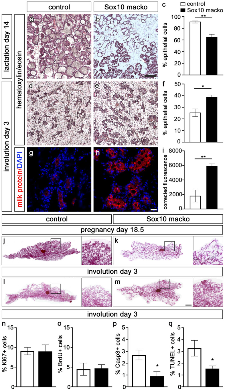
Analysis of involution of the mammary gland in Sox10 macko mice. (a–f) Hematoxylin/eosin staining of mammary gland sections was performed to compare the histology in adult control (a,d) and Sox10 macko (b,e) mice at 14 days of lactation (a–c) and 3 days of involution (d–f) and to quantify the percentage ± SEM of epithelial cells in the gland (c,f) (n = 3–5). (g–i) Immunohistochemistry with antibodies (red) directed against mouse milk proteins (g,h) was used to quantify the milk protein signal on mammary gland sections of adult control (white bars) and Sox10 macko (black bars) mice at 3 days of involution as corrected fluorescence ± SEM. (i). Nuclei were counterstained with DAPI. (n = 3–4). (j–m) Carmine-alum stainings were performed at day 18.5 of pregnancy (j,k) and day 3 of involution (l,m) on control (j,l) and Sox10 macko (k,m) mice. Magnifications on right correspond to boxed areas in main panels. Scale bars: 500 µm (b), 50 µm (h), 1 mm (m). (n–q) Quantification of the percentage of mammary epithelial cells ± SEM at day 3 of involution in control (white bars) and Sox10/tdTomato macko (black bars) mice that are labelled by antibodies directed against Ki67 (n), BrdU (o), cleaved caspase 3 (p) or by TUNEL (q) (n = 5–8). Statistical significance of differences between control and Sox10 macko was determined by Student’s t-test (*, P ≤ 0.05; **, P ≤ 0.01).
