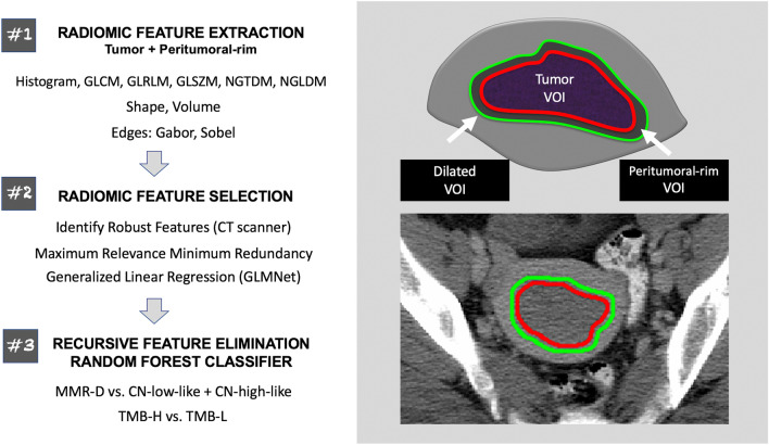
Figure 1.
Schematic overview of the methods employed in this study. The radiologist-defined tumor VOIs were used to extract intra-tumoral radiomic features. Peritumoral-rim was generated by automatically expanding the tumor contours by 3 mm and subtracting the dilated VOI from the tumor VOI. Peritumoral-rim features were computed as the differences in the values of radiomic features between the dilated VOI and the tumor VOI. The features are later down-selected via a multi-step approach; the selected features were z-score standardized and used to construct the classifiers. CT computed tomography, GLCM gray level co-occurrence matrix, GLRLM gray level run length matrix, GLSZM gray level size zone matrix, NGTDM neighborhood gray tone difference matrix, NGLDM neighborhood gray level dependence matrix, MMR-D DNA mismatch repair-deficient, CN copy number, TMB-H tumor mutational burden-high, TMB-L tumor mutational burden-low, VOI volume of interest.

