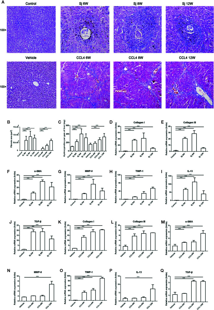
Figure 2.
The intensity of liver fibrosis and cytokine levels in the liver of mice infected with S. japonicum or injected with CCl4 intraperitoneally. (A) Masson staining of the liver in mice. Original magnification 100×. (B)The fibrosis areas of the liver of mice infected with S. japonicum or injected with CCl4 intraperitoneally. (C) The hydroxyproline level in the mice liver. (D–J) The relative mRNA expression levels of collagen I, collagen III, α-SMA, MMP-9, TIMP-1, IL-13, and TGF-β in the liver of mice infected with S. japonicum. (K–Q) The relative mRNA expression levels of collagen I, collagen III, α-SMA, MMP-9, TIMP-1, IL-13, and TGF-β in the liver of mice injected with CCl4 intraperitoneally. SJ, S. japonicum Vehicle, olive oil. Data represent the mean ± SE from three independent experiments. *** P < 0.001.

