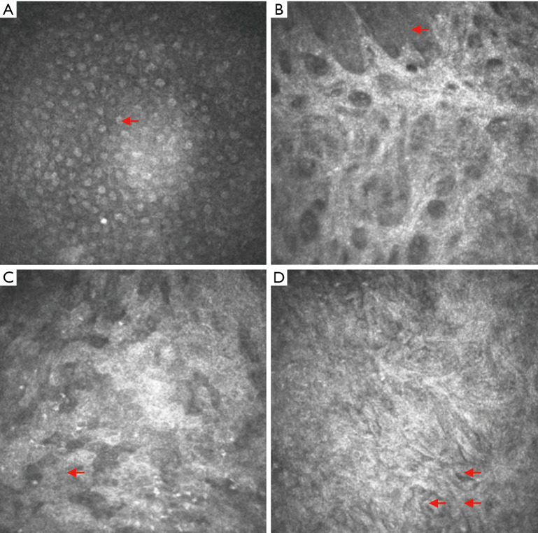
Figure 5.
In vivo dynamic observation of the corneal epithelium using confocal corneal microscopy. (A) Normal corneal epithelium. The red arrows point to the cornea, with low-reflection cytoplasm and a high-reflection nucleus. (B) Corneal epithelial defects. The exposed collagen matrix layer can be directly observed. The red arrow points to the remaining corneal epithelial cells. (C) Example from the amniotic membrane transplantation group. A large number of irregular cells can be seen. The arrow points to inflammatory cells. (D) Example from the rADSC combined with amniotic membrane transplantation group. The arrows point to spindle-shaped, tightly arranged cells. rADSC, rabbit adipose tissue-derived stem cell.

